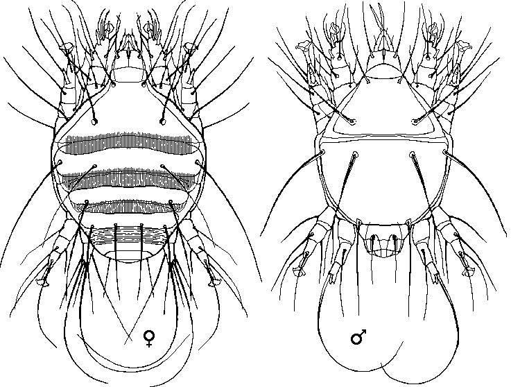
Acarine mites are parasites that invade the trachea of honey bees, but they are not much of a nuisance to beekeepers in the UK at this moment.
At large magnification they are ugly looking beasts...

This diagram has been produced as an average of several old drawings of the dorsal views of adult mites, and is similar to (inspired by) one drawn in 1921 by Stan Hirst.
In many texts the acarine mite is considered the causal agent of "Isle of Wight Disease". I have no doubt that it had an effect in that case, but I strongly doubt that it was any more than a vector or catalyst.
It was not identified until November 1920*(see below), when it was observed and named by Dr. J. Rennie (It was named Tarsonemus Woodii in the first instance and later reclassified in 1921 by Stanley Hirst).
Field Identification can be carried out with a hand lens, but even if a lens is not available it is possible to make a diagnosis with the unaided eye. The method is simple, remove the head and peel off the collar (tweezers may help). Normal thoracic tracheae are clear, colourless, or pale yellow in colour. If one or the other of the two main trachea are coloured orange or have light brown patches, then infestation is definite. Infested tracheae have patches of red/brown discolouration if the infestation is slight, but the patches will be larger and appear dark brown or even black in heavier infestations. If the trachea are white then the bee is either free of the parasite or the infestation is in the very earliest stages.
Microscopic examination using a binocular dissecting type of instrument with a magnification anywhere in the range 10x to 60x is possible, but 15x or 20x will be the most comfortable. The method of working is explained on the Diagnosis of disease and Acarine Diagnosis pages.
Life Cycle
Most the life cycle of Acarapis woodi(i) is spent within the trachea of adult honey bees where it reproduces and feeds. There is a brief time in which migration of pregnant female mites can occur. Much has been stipulated about the duration of this time and the stiffness of hairs that protect the entrance to the first thoracic spiracle (generally accepted as being up to 24 hours after emergence of the bee). I have, strong personally held, views that the length, stiffness and hardening rate of these hairs may vary from strain to strain and as a result more investigation needs to be carried out to determine the fine details.
The mites penetrate the breathing tube walls with their mouth parts and feed on the Haemolymph. About 3 or 4 days after entry a female mite starts to lay eggs. Eggs hatch in 3 to 4 days, larval form of the mite has only six legs, as opposed to the eight legs of the adult stage.
Mites are spread throughout a colony as a result of direct physical contact between older, infested bees and young bees that are as yet un-affected.
Mites have been occasionally found in air sacs in the thorax and abdomen.
Mites leave the trachea after the death of the bee.
* According to Manley... the acarine mite was first found by Miss E. Harvey, in December 1919. The research was first conducted Dr. Rennie and financed by Mr. Wood, in memory of whom the species was named Acarapis woodi. The report was published in 1921 by the Royal Society of Edinburgh in a large, illustrated pamphlet. As Miss Harvey was mentioned by name and there may have been some manipulation at the time, for whatever reason, it is quite possible this is the more accurate account.